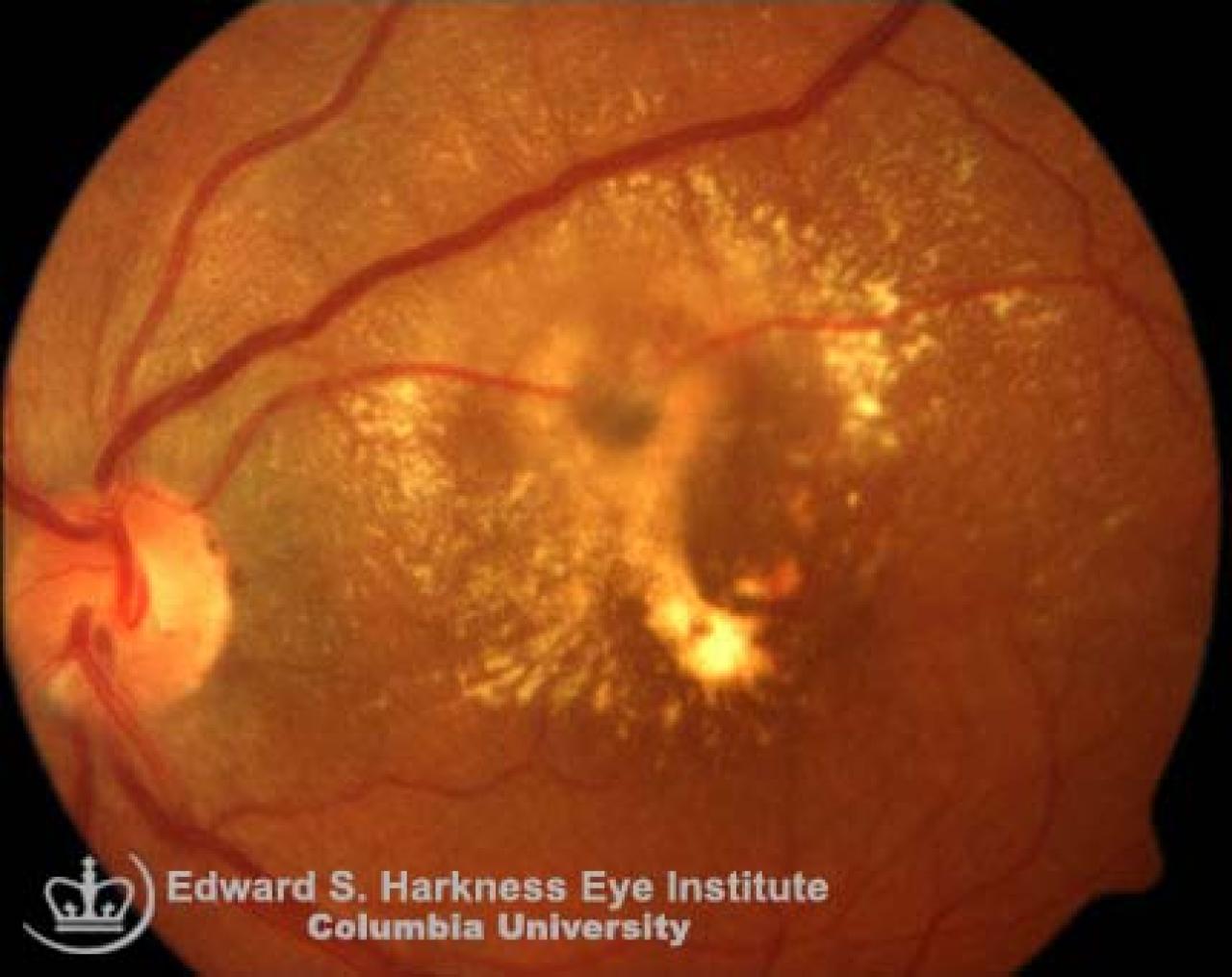
Retinal Artery Macroaneurysm
- Aquired retinal vascular abnormalities.
- Mostly affects women.
- Most commonly involves supratemporal artery.
- Associated with systemic arterial hypertension in about 60% of cases.
Clinical Features
- Symptoms:
- Maybe asymptomatic
- Decreased visual acuity if retinal edema, exudation and hourglass hemorrhages involving the macular area
- Signs:
- Round or fusiform dilations of retinal arteries within the first three orders of arteriolar bifurcation
- Usually unilateral
- Occasionally multiple
- Retinal edema
- Circinate configuration of lipoprotein exudation in the nerve fiber layer
- May present with hemorrhages into the retinal, subretinal space or vitreous cavity (hourglass hemorrhages)
- Fluorescence angiography typically demonstrates:
- Early hyperfluorescence
- Staining of the vessel wall or various degree of dye leakage in the late phase
- The aneurysm may not be visible due to the blockage of associated hemorrhage and/or hard exudate
- Management with laser photocoagulation in selected cases.