Skip to content
Cystoid Macular Edema
- One of the most common cause of vision loss following uncomplicated cataract removal either with or without implantation of intraocular lens
- Other conditions that may be complicated with CME include: diabetes, intraocular inflammation, vascular occlusions, epiretinal membrane, macroaneurysm, exudative age-related macular degeneration, hypotony and retinal detachment
Clinical Features
- Symptoms:
- Reduced visual acuity
- Hyperopic shift refraction
- Signs:
- Loss of foveal depression
- Thickening of the retina associated with translucent intraretinal cystoid spaces at the posterior pole
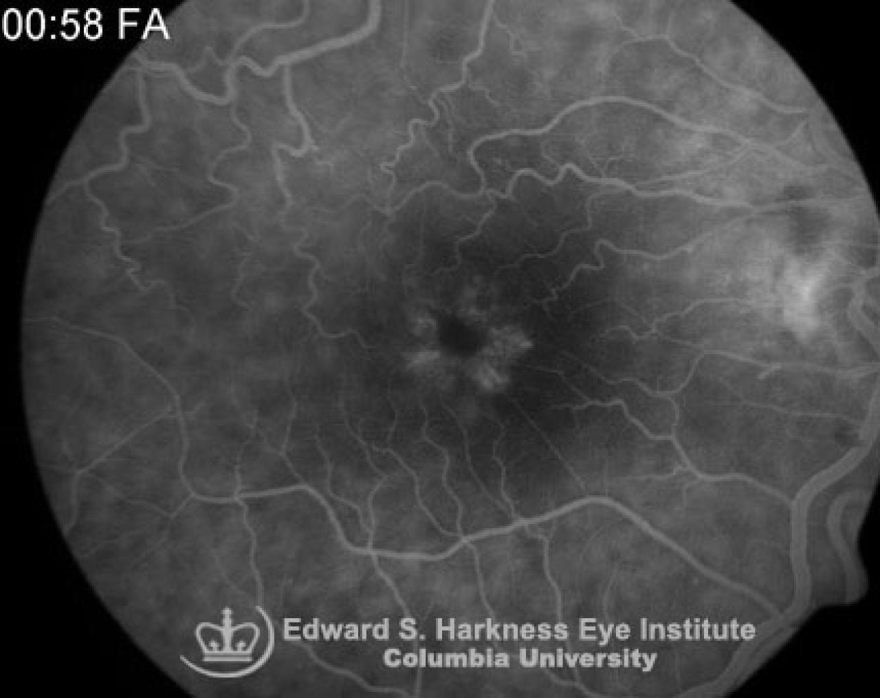
Fluorescein angiography demonstrates:
- Dye leakage from small points in the midsection of each capillaries
- Pooling of fluorescein in obliquely oriented henle layer which gives rise a characteristic petaloid staining patter in the perifoveal region
- Late staining of the optic nerve is associated with inflammatory CME, typically after cataract extraction
- Optical coherence tomography (OCT) is very helpful for diagnosis as well as for follow-up of treatment
Management
- Rule out infectious process, intraocular derangement such as entrapment of the iris or vitreous prolapse in the wound, uveitis or diabetic retinopathy
- Therapeutic approach with topical corticosteroid or cyclo-oxygenase inhibitor
- Sub-tenon's or intravitreal corticosteroid injection in refractory cases
- Nd:YAG laser vitreolysis
- Vitrectomy in selected cases
- Intraocular lens removal or replacement
Back to top