Skip to content
Limbal Dermoid
Clinical Features
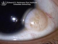
- Pale yellowish solid mass lesions, which frequently contain hair shafts
- Most often located at the lower temporal limbus involving conjunctiva and cornea
- Contain cellular elements from ectodermal and mesodermal origin such as hair follicles, sebaceous and sweat glands, ectopic lacrimal gland and cartilage
- Often associated with other congenital ocular and systemic abnormalities such as oculoauriculovertebral dysplasia (Goldenhar syndrome), Duane's syndrome, coloboma of the upper lid, and lacrimal stenosis.
Management
- Solid mass removal may be performed for cosmetic reasons or if the mass encroaches towards the central cornea resulting in astigmatism and visual impairment.
- Tumor excision at the level of corneal surface may be done to remove the mass with less posterior extension.
Back to top
