Skip to content
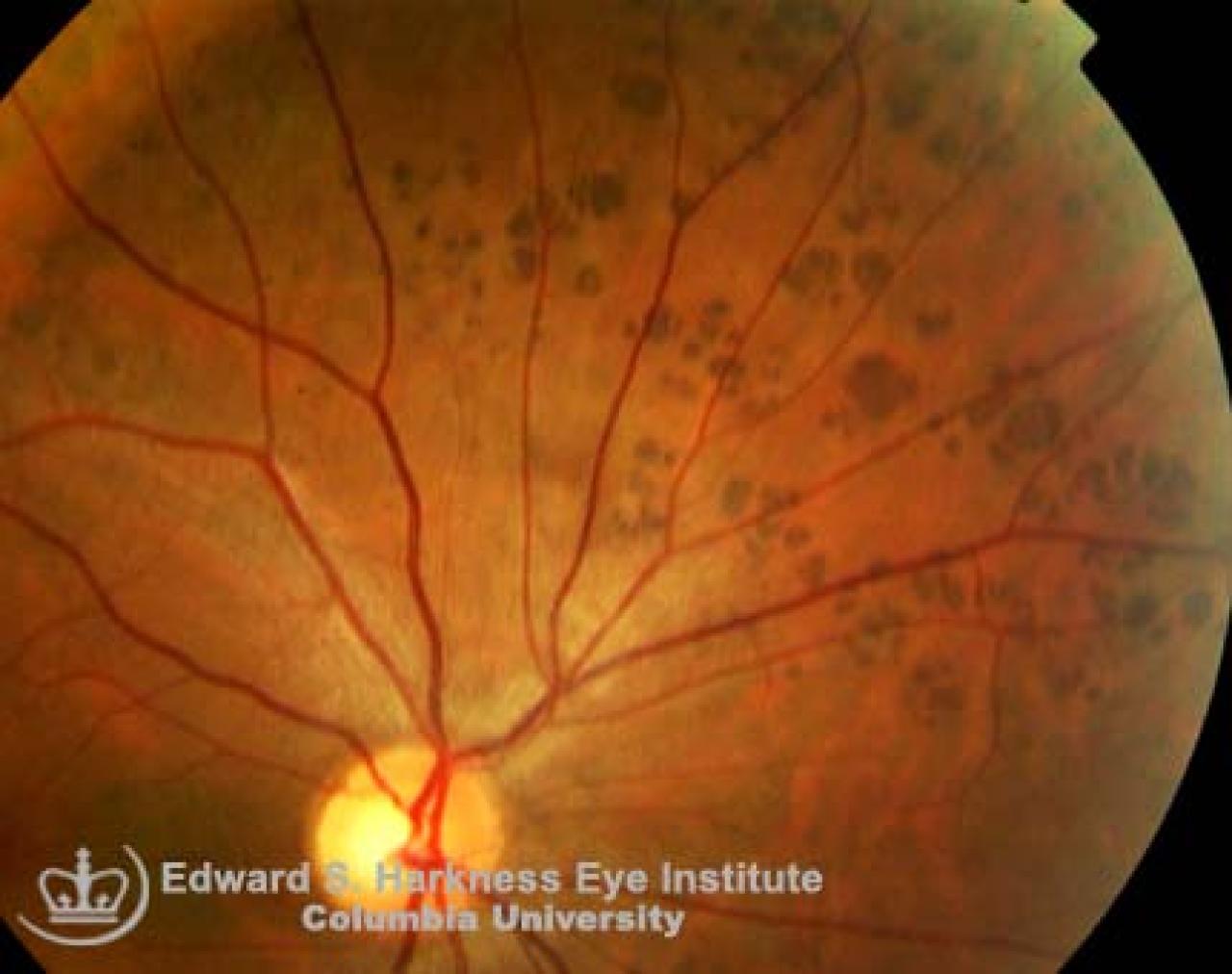
"Bear Tracks" CHRPE
- Benign pigmented fundus lesions that commonly discovered during routine eye examination.
Clinical Features
- Usually asymptomatic.
- Signs:
- Well-demarcated, round, solitary or multiple gray-brown or black lesions which have flat or scalloped margins.
- May be encircled by hyper- or hypo-pigmented halo.
- Depigmented or hypopigmented punched-out lacunae or fenestration lesions may be evident within larger lesions.
- Multiple areas of grouped CHRPE simulating the animal foot-print are also called "bear tracks".
- Generally located in the peripheral but may occasionally in the peripapillary region.
- Fluorescein angiography demonstrates blocked choroidal fluorescence by the hypertrophied RPE and no leakage of dye.
- Differential diagnosis include: choroidal melanomas, choroidal nevi, melanocytomas of the choroids, hyperplasia of the RPE, post-hemorrhage hemosiderin deposits.
- Known to be associated with other systemic findings such as familial adenomatous polyposis and Gardner's syndrome (intestinal polyposis, hamartoma of the skeleton, and multiple soft tissue tumors).
Back to top