Dr. Silverman has been involved in ultrasound research in ophthalmology for over 30 years. His research includes development of high-resolution imaging systems, studies of ultrasound safety and bioeffects, high-intensity ultrasound, blood-flow imaging, photoacoustics, and tissue characterization by use of signal-processing. He applies these techniques for studies of ocular disease in animal models and for clinical examinations.
Dr. Silverman is currently Principle Investigator on an NIH-sponsored project whose goal is development of a novel ultrasonic imaging technique, ultrafast plane-wave imaging, which enables acquisition of up to 10,000 images per second. Computer-analysis of the data allows visualization and measurement of blood-flow throughout the eye and orbit. This technique is being applied to glaucoma, vascular malformations and occlusions, and retinopathy of prematurity. Technical collaborators on this project include Raksha Urs, PhD, Jeffrey Ketterling, PhD (Riverside Research) and Alfred Yu, PhD (University of Waterloo).
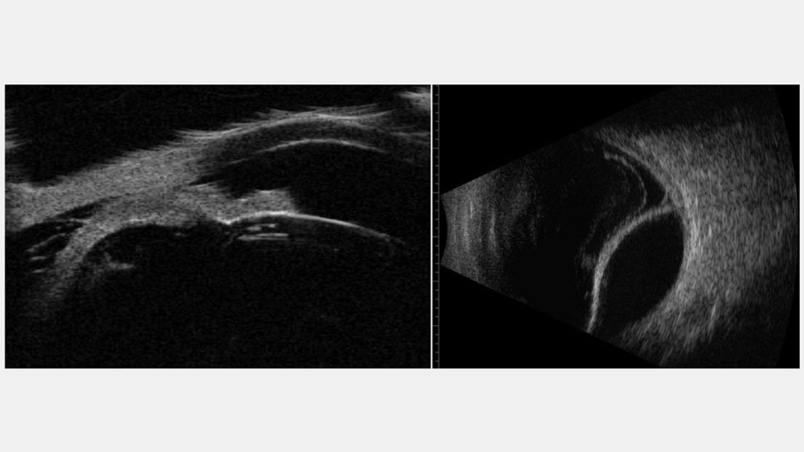
Diagnostic ultrasound images of a 69 year old male with hypotony
UBM image (left) reveals ciliary body effusion. 10 MHz B-scan (right) demonstrates choroidal detachment.
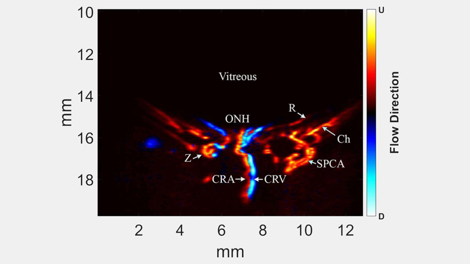
Plane-wave ultrasound Doppler image of posterior pole of pre-term neonate
ONH=optic nerve head; CRA=central retinal artery; CRV=central retinal vein; SPCA=short posterior ciliary artery; Ch=choroidal artery; R=retina, Z=circle of Zinn.
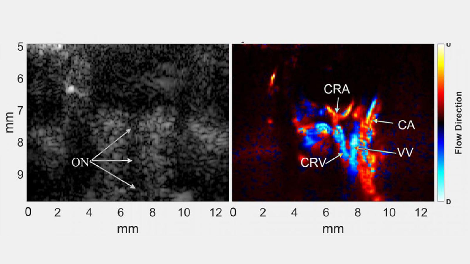
Plane-wave B-mode image (left) and corresponding plane-wave
Doppler image of posterior pole and orbit of a rat eye after intravenous injection of microbubble contrast agent. ON=optic nerve; CRA=central retinal artery; CRV=central retinal vein; VV=vortex vein; CA=ciliary artery.
Diagnostic ultrasound images of a 69 year old male with hypotony
UBM image (left) reveals ciliary body effusion. 10 MHz B-scan (right) demonstrates choroidal detachment.