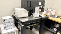
Microscopy Core

The Microscopy core of the Columbia Center for Translational Immunology (CCTI) provides training and access to state-of-the-art microscopes for biomedical investigators at the Columbia University Irving Medical Center. This core is particularly useful for imaging, qualitative analysis, and quantification of cells.
Please contact the facility staff for any issues regarding training, assistance and access including sample preparation, protocol design, instrument operation, data analysis, and troubleshooting.
Instruments
Zeiss 900 Confocal Laser Scanning Microscope
- Two new sensitive multi alkali PMT detectors
- Four lasers: 405, 488, 561, and 640 nm
- Motorized stage.
- Equipped for live cell imaging: On stage incubator with control of temperature, CO2 and humidity.
- ZEN Blue software
- Location: BB 17-1731A
Zeiss Axioimager A2 LED Microscope
- Manual stage
- Axiocam 503 camera
- Excite LED 120 automated illumination system
- Equipped with 4 filters, DAPI, 488, 546 and far red
- Five achroplan objectives
- ZEN 2 Pro software
- Location: BB 17-1731A
Fees
$45 per one hour of self-use, $100 per one hour of training and supersized sessions.
Use of Equipment
Please email the following to discuss which instrument best fits the experiment and training will be arranged accordingly. Please contact the core for generating the online instrument reservation account.
- Adam Mor
am5121@cumc.columbia.edu - Syed M. Bukhari
smb2326@cumc.columbia.edu
Reservations
All reservations are made through iLab.
Data Policy
The core does not backup any data, and to facilitate the smooth operation of the microscopes, deletes all files and templates frequently without prior notice. We recommend to save all data to your network share or OneDrive.
Getting Started
- Web-based theory session (1.5 hours) covers the principle of confocal imaging.
- The hands-on training session (2 hours) includes the instrument startup/shutdown, software usage, data storage, and troubleshooting.
